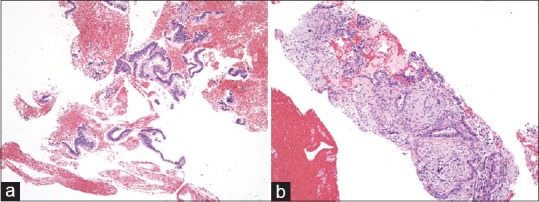
Figure 4.

(a) A small amount of fragmented adenocarcinoma cell clusters obtained using a conventional end-cut type needle (H and E, ×100), which is difficult to differentiate from contaminated gastric foveolar epithelium. The evaluation of invasive growth is impossible based on this section. (b) A core tissue including the desmoplastic fibrosis with neoplastic cellular elements obtained using a novel Franseen needle (H and E, ×100). Destructive invasion growth is apparent, leading to an accurate diagnosis for malignancy
