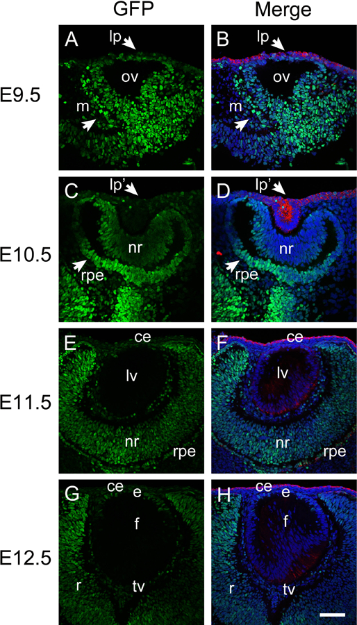
Figure 1.

Activation of canonical Wnt signaling (revealed by GFP expression) during early embryonic eye development. Immunofluorescent staining for GFP (green) and keratin 8 (red) in E9.5 (A-B), E10.5 (C-D), E11.5 (E-F) and E12.5 (G-H) embryonic mouse eye. Panels A, C, E, and G, are GFP only, panel B, D, F, and H are merged images. Blue-DNA, Green-GFP, Red-keratin 8, lp, lens placode; ov, optic vesicle; m, periocular mesenchyme; lp, lens placode; lp’, lens pit; nr, neuroretina; rpe, presumptive retinal pigmented epithelium; ce, cornea epithelium; lv, lens vesicle; e, lens epithelium; f, lens fibers; tv, tunica vasculosa; scale bar = 70μm.
