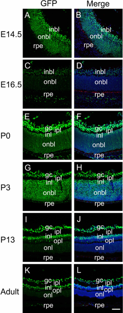
Figure 4.

The distribution of canonical Wnt signaling (represented by GFP) during retina development. Immunofluorescent staining for GFP (green) and keratin 8 (red) in E14.5 (A-B), E16.5 (C-D), P0 (E-F), P3 (G-H), P13 (I-J) and adult (K-L) mouse retina. Panels A, C, E, G, I and K are GFP plus keratin 8, panels B, D, F, H, J and L are merged images including the DNA channel. Blue-DNA, Green-GFP, Red-keratin 8; inbl, inner neuroblastic layer; onbl, outer neuroblastic layer; Gc, ganglion cell; rpe, retinal pigmented epithelium; ipl, inner plexiform layer; inl, inner nuclear layer; opl, outer plexiform layer; onl, outer nuclear layer; scale bar = 70μm.
