
Figure 5.
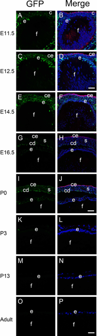
The distribution of canonical Wnt signaling (represented by GFP) during lens development. Immunofluorescent staining for GFP (green) and keratin 8 (red) in E11.5 (A-B), E12.5 (C-D), E14.5 (E-F), E16.5 (G-H), P0 (I-J), P3 (K-L), P13 (M-N) and adult (O-P) mouse lens. Panels A, C, E, G, I, K, M and O are GFP only, panels B, D, F, H, J, L, N and P are merged images. c-cornea; l-lens; ce-cornea epithelial cells; e-lens epithelial cells; f-fiber cells; s-cornea stroma; cd-cornea endothelial cells. Blue-DNA, Green- GFP, Red- keratin 8, A-D and K-P, scale bar = 35μm, E-J, scale bar = 70μm.