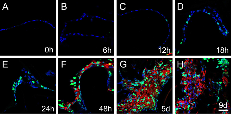
Figure 6.

The activation of canonical Wnt signaling (represented by GFP) in lens epithelial cells remaining behind in a mouse model of cataract surgery. Immunofluorescent staining for GFP (green) and αSMA (red) in 0h, 6h, 12h, 18, 24h, 48h, 5 days and 9 days post-surgery samples. A) GFP expression was not detected at 0h post-surgery, B) GFP expression was not detected at 6h post-surgery, C) GFP expression was detected in occasional lens epithelial cell nuclei at 12h post-surgery, D) GFP expression becomes more prevalent in lens epithelial cells at 18h post-surgery, E) GFP expression continues to upregulate at 24h post-surgery, F) robust GFP and αSMA expression co-localize in remnant lens cells at 48h post-surgery, G) robust GFP and αSMA expression co-localize in remnant lens cells at 5 days post-surgery, H) robust GFP and αSMA expression persist in remnant lens cells at 9 days post-surgery. Blue-DNA, Green-GFP, Red-αSMA, scale bar = 35μm
