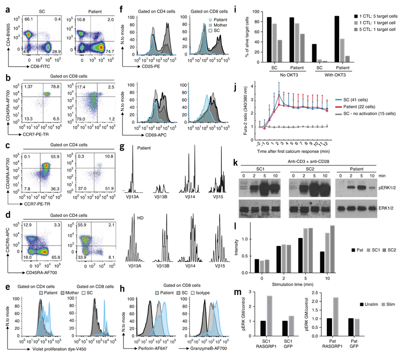
Figure 2. RASGRP1deficiency causes defective TCR signaling and an aberrant immunophenotype.
(a–d) T cell immunophenotyping displaying relative proportions of CD4+ cells and CD8+ cells amongst CD3+ cells in lymphocyte gate (a), proportions of naive (CD45RA+CCR7+), central memory (CD45RA−CCR7+)T cells, effector memory (CD45RA−CCR7−) T cells and exhausted effector memory (CD45RA+CCR7−) T cells among CD8+ T cells (b) and CD4+ T cells (c), and the relative abundance of CXCR5+CD45RA− cells and naive (CXCR5−CD45RA+) cells among CD4+ cells (d). Full gating strategy, Supplementary Figure 3. (e) Proliferation of CD4+ and CD8+ T cells at day 4 after stimulation with anti-CD3 and anti-CD28. (f) Expression of CD25 and CD69 24 h after stimulation with anti-CD3 and anti-CD28. (g) TCR Vβ spectra-typing of the patient’s peripheral blood T cells. (h) Expression of perforin and granzyme B on the patient’s CD8+ T cells. (i) Killing of target cells by CD8+ T cells with (right) or without (left) OKT3. CTL, cytotoxic T lymphocytes. (j) Microscopy of intracellular Ca2+ flux in immortalized T cell lines following stimulation with anti-CD3, assessed by detection of the Fura-2 excitation ratio. (k) Cropped immunoblot analysis of total and phosphorylated (p-) ERK in primary T cells after stimulation with anti-CD3 and anti-CD28. (l) Quantification of the results in k. (m) Geometric mean (GM) of phosphorylated ERK in the patient’s CD8+ T cells and shipment control CD8+ T cells transfected to express a vector encoding wild-type RASGRP1 or a vector encoding green fluorescent protein (GFP) as control. Data are representative of eight (a), four (b,c), three (e,f,h,i,m) or two (d,g,j,k) independent experiments.

