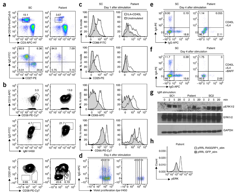
Figure 3. RASGRP1 deficiency results in a B cell proliferation and activation defect.
(a) Showing relative proportion of naive (IgD+CD27−) B cells, memory non-switched (IgD+CD27+) B cells and memory-switched (IgD−CD27+) B cells. Full gating strategy, Supplementary Figure 3. (b) Flow cytometry of gated CD19+ B cells showing transitional T1 (CD10+CD38+) B cells, transitional (IgD+IgM+) B cells and activated (CD21−CD38lo) B cells. (c) Flow cytometric expression of CD86, CD95, CD25 and CD69 of CD19+ cells 1 d after stimulation with anti-CD40L and IL-4. (d) Proliferating B cells at day 4 after stimulation as in c. (e) IgA+ and IgG+ switched B cells at day 4 stimulation. (f) IgA+ and IgG+ switched B cells at day 4 stimulation as in c with (bottom) or without (top) the addition of BAFF. (g) Cropped immunoblot analysis of total and phosphorylated ERK in EBV-transformed B cells from the patient, assessed after stimulation with IgM. (h) Phosphorylated ERK after gene transfer using a lentiviral backbone (pRRL) with sequence encoding wild-type RASGRP1 or GFP in EBV-transformed B cells from the patient, after stimulation with IgM. Data are representative of four (a,b), two (c,d,g,h) or one (e,f) independent experiment(s).

