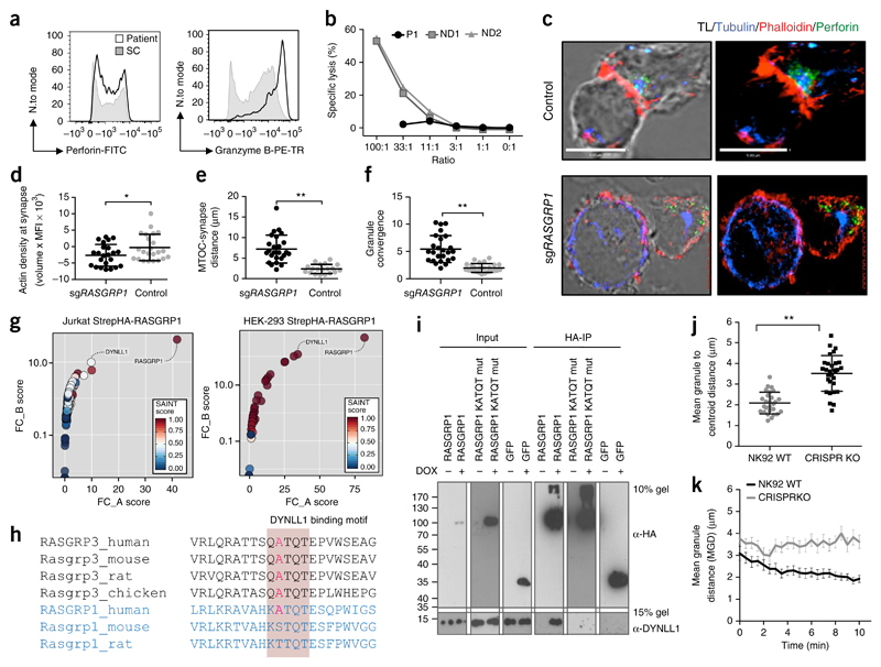
Figure 4. Aberrant cytoskeletal dynamics in NK cells.
(a) Flow cytometry analyzing the intracellular accumulation of perforin and granzyme B in NK cells from the patient and control donors. Full gating strategy, Supplementary Figure 4. (b) Chromium (51Cr)-release assay of NK cell cytotoxicity. E/T, effector cell/target cell. (c) Confocal microscopy of a fixed RASGRP1 wild-type NK-92 control cell (top) and an NK-92 cell with CRISPR-mediated editing of RASGRP1 (sgRASGRP1) (bottom) conjugated to K562 cells, detected with anti-α-tubulin (blue), anti-perforin (green) and phalloidin-stained F-actin (red). TL, transmitted light. (d–f) Actin density at the synapse (d), distance from lytic granule to MTOC (e), and granule convergence to MTOC (f) of RASGRP1 wild-type NK-92 control cells and sgRASGRP1 NK-92 cells. (g) Tandem affinity purification of RASGRP1 in Jurkat cells (left) and HEK cells (right), presented as FC-A scores versus FC-B scores. (h) Conservation of the KATQT motif in RASGRP1 and RASGRP3 across various vertebrate species. (i) HA-based co-immunoprecipitation of Strep-HA–tagged wild-type (WT) RASGRP1, KATQT-mutant (mut) RASGRP1 or Strep-HA–tagged-GFP with endogenous DYNLL1. (j,k) Video microscopy of granule convergence in sgRAGSRP1 and control NK cells. *P < 0.05 and **P < 0.0001 (unpaired Student’s t test). Data are representative of four (c), three (a,c–f,i), two (g,j,k) or one (b) independent experiment(s) (mean and s.d. in d–f,j,k).

