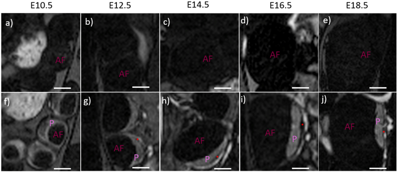
Figure 2:
T1-weighted pre and post-contrast images of individual feto-placental units demonstrate visibility of the retroplacental clear space (RPCS). (a-e) T1-weighted images without contrast have very low signal within the feto-placental unit. The placenta (P) and amniotic fluid (AF) of the fetal compartment are indistinguishable at all five gestational timepoints. (f-j) High signal is seen in the placenta and feto-placental vasculature in post-contrast T1-weighted images of the same animals. The RPCS (red asterisk) is visible starting on day 12.5 and is present at successive time points. Scale bars in each figure represent 3 mm.

