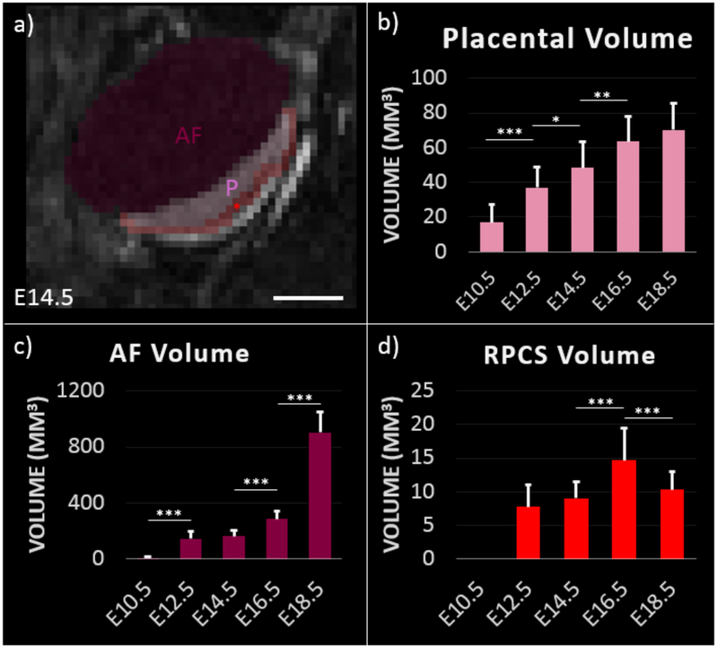
Figure 4:
Segmentation of contrast-enhanced T1-weighted MRI enables longitudinal monitoring of feto-placental development. a) Segmentation of feto-placental unit at day 14.5 of gestation. Placenta (P), amniotic fluid (AF), and RPCS (red asterisk) are labeled. Scale bar represents 3 mm. Volume estimates of b) placenta, c) amniotic fluid, and d) retroplacental clear space at each gestational time point are. Error bars indicate standard deviation among fetal placental units at each time point. Significance as determined by the Wilcoxon rank sum test are shown for p<0.05 (*), p<0.005 (**), and p<0.0005 (***).

