
FIG 2. IgG+ B cells from CVID+AIC patients display low SHM frequencies and an altered VH repertoire.
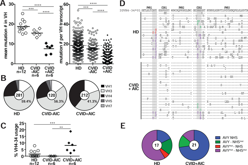
(A) Mutations in VH transcripts from CD27+IgG+ B cells from 12 healthy donors (HDs, open circles), 6 CVID patients without AICs (CVID-AIC, open diamonds) and 6 CVID+AIC patients (black diamonds) are displayed as averaged mutation number per subject (left) and absolute mutation number per transcript (right). Black bars represent mean values. (B) Pie charts represent VH family gene segment usage from pooled IgG transcripts. Transcript numbers per group are indicated in each pie’s center. (C) Averages of VH4-34gene segment usage in IgG+ B cells. (D) VH4-34 amino acid sequence alignment for IgG+ memory B cells from HDs and CVID+AIC patients relative to germline IGHV4-34*01 (top). Identity to germline is denoted by a dash and substituted residues are indicated in black unless within the critical AVY or NHS motifs (gray columns) where text color is red or green, respectively. Substitutions within both motifs are indicated in purple. (E) Pie charts represent proportions of pooled transcripts in HDs and CVID+AIC patients with AVY and/or NHS substitutions. Statistically significant differences are indicated: ****P <0.0001, ***P <0.001, **P <0.01 (Mann-Whitney U tests).