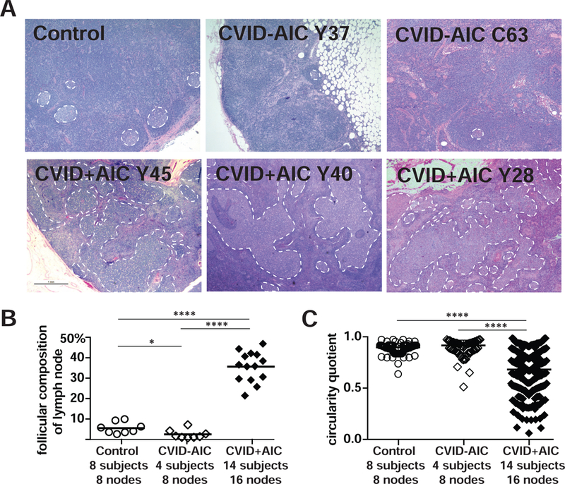
FIG 3. Asymmetric enlarged germinal centers (GCs) predominate in lymph nodes from CVID+AIC patients.

(A) Hematoxylin- and eosin-stained axillary lymph node biopsies from a representative immunocompetent 43-year-old female (Control), two CVID-AIC patients, and three CVID+AIC patients. GCs are outlined with white dashed lines. Original magnification, 12.5X. (B) The percentages of each lymph node’s cellular two-dimensional surface comprised by GCs and (C) each GCs circularity quotient are represented for controls and patients. Statistically significant differences are indicated: ****P <0.0001, **P <0.01 (Mann-Whitney U tests).
