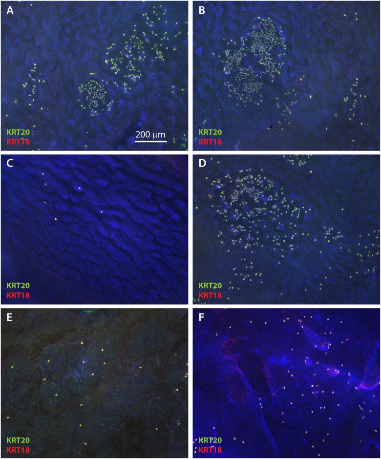
Fig 3. Merkel cell density in ESS is highly variable.
Shown are en face images of epidermis from ESS at 12 weeks (A-B) and 14 weeks (C-D) after grafting, and normal human skin (E-F), immunostained with antibodies against KRT20 (green) and KRT18 (red); DAPI was used to counterstain nuclei (blue). Tissue was photographed at low magnification to illustrate variability in density of Merkel cells and relatively random distribution. Scale bar in A (200 μm) is same for all panels.

