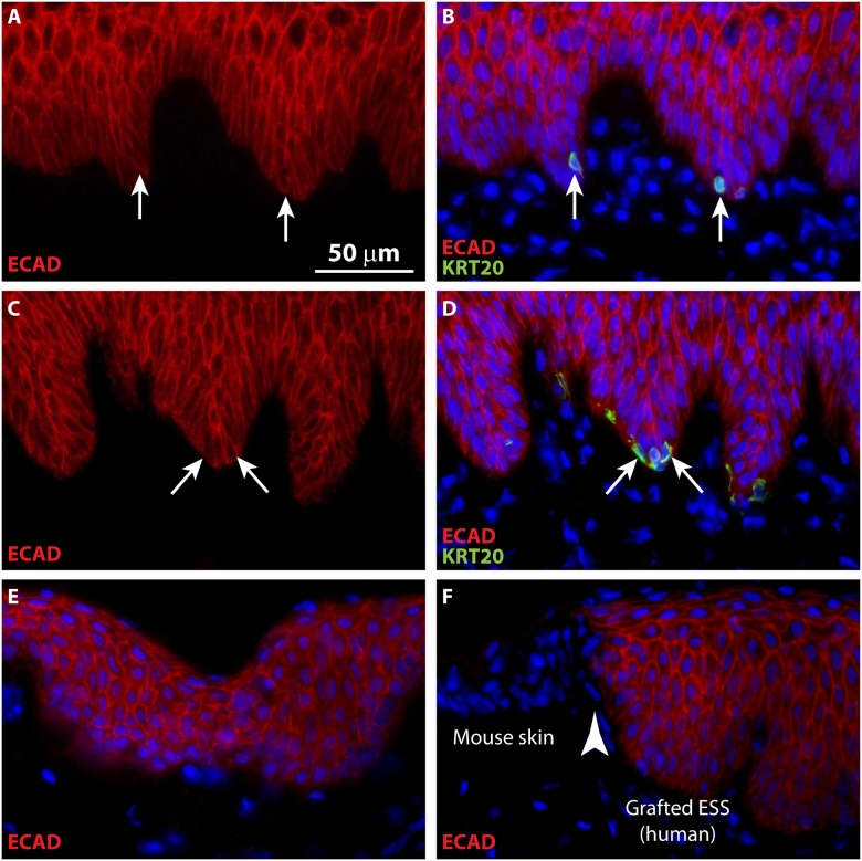
Fig 5. Merkel cells in grafted ESS are derived from human epidermal cells.
Immunohistochemistry was performed using a human-specific antibody against E-cadherin (ECAD; red); Merkel cells were localized by KRT20 immunostaining (green). A-B, ESS at 6 weeks after grafting; C-D, ESS at 12 weeks after grafting. Sections are oriented with the epidermis at the top of each image; in A-D, each row depicts images of the same tissue section. Arrows depict examples of KRT20-positive Merkel cells in ESS. E-F, Controls for native human skin (E) and immunodeficient mouse skin (F) demonstrating specificity of anti-human E-cadherin antibody (red). Section shown in F depicts the border (arrowhead) between grafted human ESS (right) and flanking mouse skin (left). Nuclei were counterstained with DAPI (B, D, E, F; blue); scale bar in A is for all sections (50 μm).

