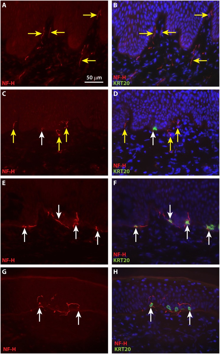
Fig 9. Merkel cells in grafted ESS are associated with neurons expression neurofilament heavy (NF-M) by eight weeks after grafting.
Immunochemistry with antibodies against NF-H (red) and KRT20 (green) was used to localize neurons and Merkel cells, respectively, in ESS after grafting to mice. Nuclei were counterstained with DAPI (blue; B, D, F, H). Shown are cross sections of ESS at 4 weeks (A-B), 8 weeks (C-D), 12 weeks (E-F), and 14 weeks (G-H) after grafting; each row contains images of the same section. Scale bars in A is same for all images. White arrows indicate examples of NF-H-positive nerves associated with or in proximity to Merkel cells; yellow arrows indicate NF-H-positive nerves not associated with Merkel cells.

