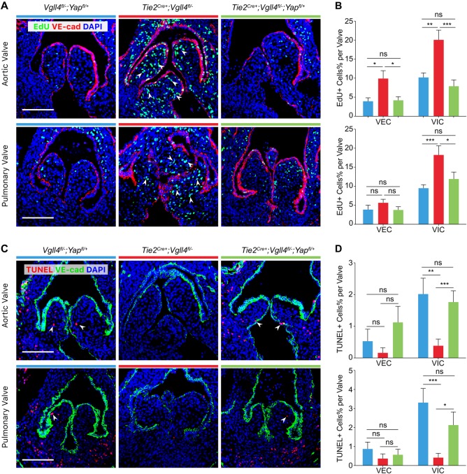
Fig 7. VGLL4 negatively regulates proliferation and promotes apoptosis of VICs during development of arterial valves.
(A) EdU staining shows proliferating cells (green) in arterial valves of E15.5 Vgll4fl/-;Yapfl/+; Tie2Cre+;Vgll4fl/- and Tie2Cre+;Vgll4fl/-;Yapfl/+ embryos. VE-cad staining marks VECs (red). (B) Quantitative results of EdU+ cells of VECs and VICs in each individual leaflet (n = 5). (C) TUNEL assay shows apoptotic cells (red) in arterial valves of E15.5 Vgll4fl/-;Yapfl/+; Tie2Cre+;Vgll4fl/- and Tie2Cre+;Vgll4fl/-;Yapfl/+ embryos. VE-cad staining marks VECs (green). (D) Quantitative results of TUNEL+ cells of VECs and VICs in each individual leaflet (n = 5). White arrows point proliferating cells (in A) or apoptotic cells (in C). VEC: valve endothelial cell; VIC: valve interstitial cell. Scale bar = 100μm. *P<0.05, **P<0.01, ***P<0.005, ns: no significance.

