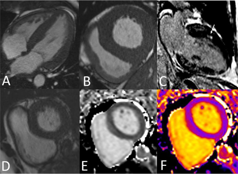
Figure 1:

CMR images of (A) Four chamber SSFP showing severe concentric LVH. (B) Basal short axis SSFP showing severe concentric LVH. (C) Two chamber view depicting anterior midwall LGE (arrow) in patient with HHD. (D) SSFP basal short axis image. (E) Pre-contrast T1 map in black & white. (F) T1 color map
