
Figure 4.
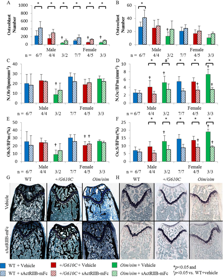
Femurs of sActRIIB-mFc treated mice demonstrated increased osteoblast number and decreased osteoclast number per bone surface and osteoclast surface per bone surface in the distal trabecular bone regardless of sex and genotype, except for male WT mice. A) Osteoblast number (N.Ob), B) Osteoclast number (N.Oc), C) Osteoblast number per bone surface (N.Ob/BPm), D) Osteoclast number per bone surface (N.Oc/BPm), E) Osteoblast surface per bone surface (Ob.S/BPm), and F) Osteoclast surface per bone surface (Oc.S/BPm) of male and female WT, +/G610C, and oim/oim vehicle (solid color) and sActRIIB-mFc (diagonal) treated mice. G) and H) Representative images of Von Kossa and TRAP stained longitudinal trabecular sections of female mice. (WT [blue], +/G610C [red], and oim/oim [green]). (n) is indicated for each genotype and treatment. The number of samples are lower in +/G610C and oim/oim due to fragility of the sections and limited number of callus free femurs. Values are MEAN±SD. *p<0.05; †p<0.05 vs. WT+vehicle treated mice; a*p=0.0673.