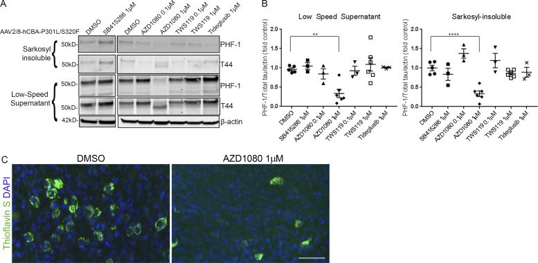
Figure 8.
Tauopathy BSCs can be used for testing tau-targeting therapeutics. (A) Representative Western blots of lysates from organotypic BSCs transduced at DIV 0 with rAAV2/8-P301L/S320F-htau (1–2 × 10¹⁰ VGs per well) and then treated from 14 to 28 DIV with 0.1 µM or 1 µM GSK-3β inhibitors. Half maximal inhibitory concentrations (IC50s) for GSK-3β for each compound are listed in parentheses as follows: SB415286 (78 nM), AZD1080 (324 nM), TWS119 (30 nM), and Tideglusib (60 nM). Blots were probed with antibodies against total tau (both nonphosphorylated and phosphorylated; 3026) and PHF-1 (phospho-Ser396/404). Blots were also probed with an antibody against β-actin as a loading control. (B) Bar charts show amounts of phospho-tau as a proportion of total tau in low speed supernatant and sarkosyl-insoluble samples after treatment with 0.1 µM or 1 µM GSK-3β inhibitors or DMSO (control). n = 3–6. **, P < 0.01; ****, P < 0.0001. Data are mean ± SEM and are shown as fold change from DMSO (control). (C) Transduced and treated slice cultures were fixed, stained with 0.0125% Thioflavin S, and imaged to identify any β-sheet structures in these sections. DAPI is also shown to mark cell nuclei. Bar, 50 µm. n = 6.

