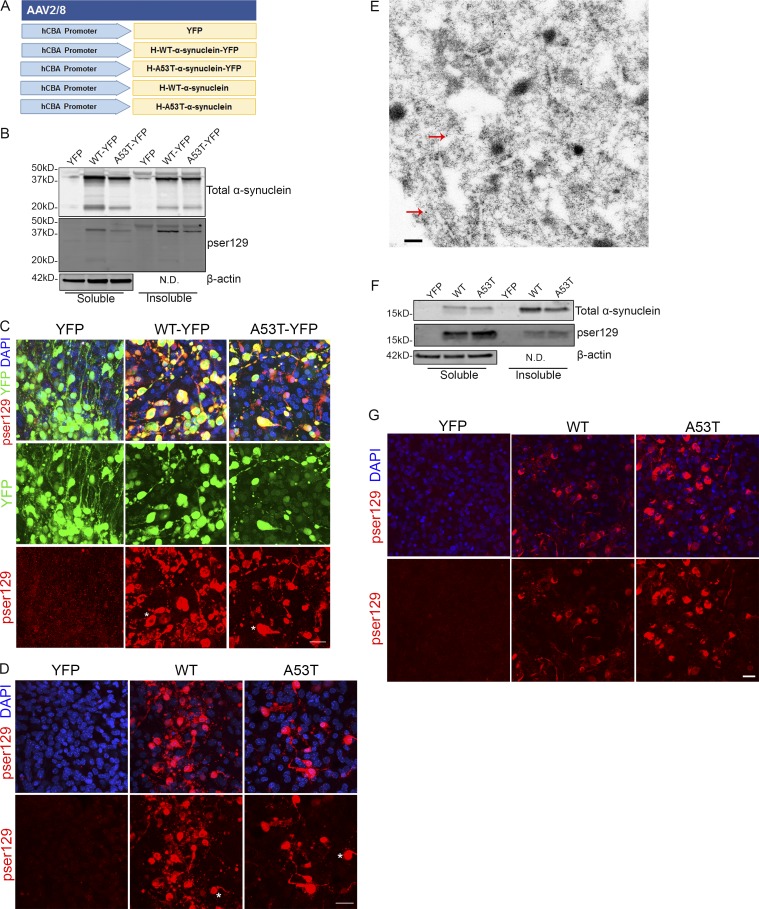
Figure 9.
rAAV α-synuclein-transduced organotypic BSCs develop insoluble α-synuclein inclusions reminiscent of LBs. (A) Schematic diagram of rAAV human mutant and WT α-synuclein constructs used to transduce organotypic BSCs. BSCs were prepared and transduced with rAAV2/8-YFP, rAAV2/8-h-WT-α-synuclein-YFP, rAAV2/8-h-A53T-α-synuclein-YFP, rAAV2/8-h-WT-α-synuclein, or rAAV2/8-h-A53T-α-synuclein prepared using a microscale method of rAAV preparation at 0 DIV and then maintained in culture until 28 DIV. (B) Transduced slices were sequentially extracted in order to prepare soluble and insoluble fractions, and then lysates were immunoblotted for total α-synuclein (SNL-4), phospho-Ser129 α-synuclein (81A), and β-actin as a loading control. Representative Western blots of insoluble and soluble fractions are shown. n = 9. (C and D) Transduced slice cultures were fixed, immunostained for phospho-Ser129 α-synuclein (EP1536Y), and confocal imaged to identify the location of phosphorylated α-synuclein in these sections. DAPI is also shown to mark cell nuclei. Asterisks mark examples of cells showing accumulation of phosphorylated α-synuclein and a LB-like appearance. Bar, 25 µm. n = 3. (E) rAAV2/8-h-WT-α-synuclein transduced slices were fixed and examined by immuno-EM for the presence of filamentous inclusions. Immunogold labeling with antibody to NACP98 confirms the presence of α-synuclein in inclusions (red arrows show examples of immunogold-labeled NACP98 on filaments). Bar, 0.2 µm. BSCs were also prepared and transduced with rAAV2/8-YFP, rAAV2/8-h-WT-α-synuclein, or rAAV2/8-h-A53T-α-synuclein prepared using the traditional purified method of rAAV preparation at 0 DIV and then maintained in culture until 28 DIV. (F) Transduced slices were sequentially extracted in order to prepare soluble and insoluble fractions, and then lysates were immunoblotted for total α-synuclein (SNL-4), phospho-Ser129 α-synuclein (81A), and β-actin as a loading control. Representative Western blots of insoluble and soluble fractions are shown. n = 3. (G) Transduced slice cultures were fixed, immunostained for phospho-Ser129 α-synuclein (EP1536Y), and confocal imaged to identify the location of phosphorylated α-synuclein in these sections. DAPI is also shown to mark cell nuclei. Bar, 20 µm. N.D., not done.

