Figure 3.
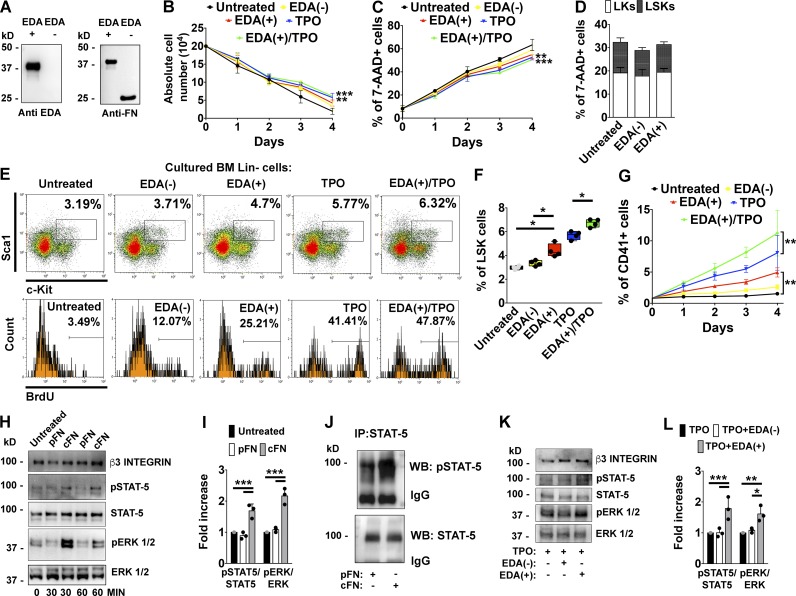
EDA domain of FN sustains hemopoietic progenitor cells proliferation and Mk development in vitro. (A) 1 µg of recombinant peptides containing (EDA+) or lacking EDA (EDA−) segment of FN were tested by Western blot with a monoclonal anti-EDA segment or a polyclonal rabbit anti-FN. (B) Lin− fractions from BM cells were maintained in DMEM for 4 d in the presence of 1 µg of EDA+ and EDA− recombinant peptides, 10 ng/ml of TPO, and TPO plus EDA+ peptide. Absolute cell numbers at different days are shown. n = 3. (C) Assessment of cell mortality by flow cytometry in the different experimental conditions using 7-AAD staining. n = 3. (D) Effects of EDA+ and EDA− peptides on LSKs and LKs survival after 24 h of culture were tested by 7-AAD staining. n = 3. (E) Upper panels: Representative flow cytometry analysis of LSK frequencies in vitro after stimulation with recombinant peptides, TPO, or TPO plus EDA+ peptide for 24 h. Lower panels: Percentage of BrdU+ proliferating cells in the LSKs gate. (F) Frequencies of LSK cells in Lin− cells stimulated with recombinant peptides, TPO, or TPO plus EDA+ peptide for 24 h. n = 4. (G) Quantification of CD41+ Mk’s at different days of culture was evaluated by flow cytometry analysis. n = 3. (H) Fetal liver–derived Mk’s were stimulated for 30 or 60 min, with 5 µg of pFN or cFN, or left untreated. STAT-5 and ERK 1/2 phosphorylation was then evaluated by Western blot. β3 integrin was revealed to ensure the same number of Mk’s used in the experiment. (I) Histograms showing the ratio of phosphorylated and total STAT-5 and ERK 1/2 proteins in Mk’s stimulated with pFN or cFN for 1 h. n = 3. (J) Mk’s were stimulated with pFN or cFN for 1 h. STAT-5 was immunoprecipitated (IP) from cell lysates and tested with a phospho–STAT-5 antibody (pSTAT-5). IgG indicates IgG chains. WB, Western blot. (K) Representative Western blot analysis of STAT-5 and ERK 1/2 phosphorylation in BM Lin− cells differentiated with TPO alone or TPO plus 1 µg of EDA− or EDA+ peptides for 4 d. (L) Histograms showing the ratio of phosphorylated and total STAT-5 and ERK 1/2 proteins in BM Lin− cells differentiated with TPO alone or TPO plus EDA− or EDA+ peptides for 4 d. n = 3. *, P < 0.05; **, P < 0.01; ***, P < 0.001. Data are shown as mean ± SD.

