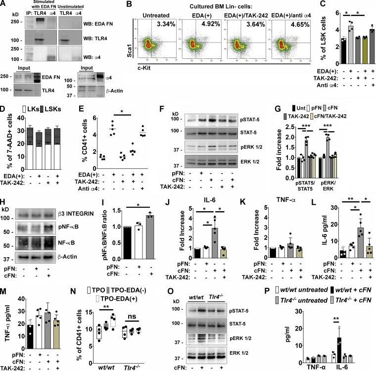
Figure 4.
Effects of EDA domain on Mk’s are TLR4 dependent. (A) Purified Mk’s were incubated for 1 h with 1 µg of cFN or left untreated, centrifuged, and lysed. Extracted proteins from both samples were split and TLR4 and α4 integrin immunoprecipitated (IP) from the two fractions of proteins, respectively. Co-immunoprecipitated cFN was then revealed in both fractions by Western blot (WB). Inputs: proteins in total cell lysates. Actin was detected from the cell lysate to ensure equal protein loading. (B and C) Representative dot plots of LSKs in Lin− cells cultured for 24 h with 1 µg of EDA+ peptide, or pretreated with TAK-242 (1 µg/ml) or an anti–α4 integrin antibody with blocking function (10 µg/ml) before EDA+ stimulation (B) and relative quantification (C). n = 4. (D) Flow cytometry analysis of 7-AAD+ dead LK and LSK cells after 24 h of culture with TAK-242 alone or in combination with EDA+ peptide. n = 3. (E) Quantification of CD41+ Mk’s derived from Lin− cells left unstimulated or stimulated with 1 µg of EDA+ peptide or pretreated with TAK-242 or an anti–α4 integrin antibody for 4 d. n = 5. (F) Purified Mk’s were left untreated or stimulated for 1 h with 5 µg of pFN, cFN, and TAK-242 alone and before cFN stimulation. STAT-5 and ERK 1/2 phosphorylation were then evaluated by Western blotting. (G) Histograms showing the ratio of phosphorylated and total STAT-5 and ERK 1/2 proteins in mature Mk’s unstimulated or stimulated with pFN, cFN, and TAK-242 alone and before cFN stimulation. n = 3. (H) Level of NF-κB activation in Mk’s stimulated with cFN for 1 h. β3 was revealed to ensure the same Mk number for each experimental condition. β-Actin was used as equal loading control. (I) Histograms showing the ratio of phosphorylated and total NF-κB in Mk’s left unstimulated or stimulated with 5 µg of pFN or cFN. n = 3. (J and K) Fold increase in IL-6 (J) and TNF-α (K) mRNA expression in Mk’s after 24 h of co-culture with pFN, cFN, and TAK-242 before cFN addition. n = 5. (L and M) IL-6 (L) and TNF-α (M) quantification by ELISA in culture supernatants from untreated and treated Mk’s. n = 5. (N) Quantification of CD41+ Mk’s derived from wt/wt and Tlr4−/− BM Lin− cells differentiated with TPO, TPO plus EDA+ peptide, or EDA− peptide. n = 4. (O) Western blot analysis of STAT-5 and ERK 1/2 activation in wt/wt and Tlr4−/− Mk’s, left untreated or stimulated with 5 µg of cFN for 1 h. n = 2. (P) TNF-α and IL-6 quantification in culture supernatants from wt/wt and Tlr4−/− Mk’s untreated, or treated with cFN. n = 3. *, P < 0.05; **, P < 0.01; ***, P < 0.001; ns, not significant. Data are shown as mean ± SD.

