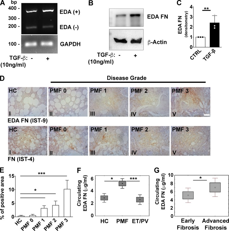
Figure 8.
Increased release of FN containing EDA domain during fibrotic progression in PMF patients. (A) Human BM-derived MSCs were stimulated for 24 h with 10 ng/ml of recombinant human TGF-β. Splicing of alternative EDA exon was analyzed by PCR assay. GAPDH was used for comparative concentration analysis. (B and C) Representative Western blot (B) and relative quantification (C) of EDA FN expression in MSCs stimulated or not with TGF-β. β-Actin was revealed to ensure equal protein loading. n = 3. Data are shown as mean ± SD. (D) Stains of FN containing EDA segment (Ab IST-9; upper panels, I–V) in BMBs of HC (upper panel I, n = 3) and PMF patients at grade 0, 1, 2, and 3 of BM fibrosis (upper panels II–V, respectively). Stains of total FN in the same specimens are shown in lower panels I–V. Bar, 100 µm. (E) EDA FN staining expressed as percent of positive area in HC and PMF at different phases of disease (n = 3 per each phase). Data are shown as mean ± SD. (F) Values of circulating EDA FN (μg/ml in 500 µg/ml of total pFN; mean ± 1.96 SE) in plasma samples of subjects stratified according the diagnosis (PMF: n = 105; PV: n = 6; ET: n = 16; or HC: n = 15). (G) Circulating plasma EDA FN (mean ± 1.96 SE) in PMF patients according to their phase of fibrotic disease: early BM fibrosis (grade 0/1, n = 30) or advanced BM fibrosis (grade 2/3, n = 8). *, P < 0.05; **, P < 0.01; ***, P < 0.001.

