Figure 4.
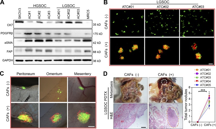
HGSOC-derived CAFs facilitate MU formation in LGSOC ATCs and promote peritoneal metastasis. (A) Immunoblotting of CAF markers (FAP, α-SMA, and PDGFRβ) and the epithelial marker CK7 in ascites cells (ACs) from HGSOC (AC#1–3) and LGSOC (AC#01–03) patients. SKOV3 served as the positive control for epithelial cells while MRC5 cells served as the positive control for fibroblasts. (B) Representative images of heterotypic spheroids formation by ATCs derived from LGSOC in suspended culture, in the absence or presence of HGSOC-derived CAFs (#2, 3, and 6). Bar, 50 µm. (C) Representative images for peritoneal adhesion of LGSOC ATCs (#02) in the absence or presence of CAFs (#2, 4, and 7) derived from HGSOC. Bar, 100 µm. (D) Representative images and quantification of peritoneal metastases in mice bearing ATCs derived from LGSOC (#01–05), in the absence or presence of HGSOC-derived CAFs (#2, 7, and 8; n = 8 mice per group). Bar, 200 µm. Data are means ± SEM and are representative of two (A, C, and D) or three (B) independent experiments. ***, P < 0.001, determined by Student’s t test.

