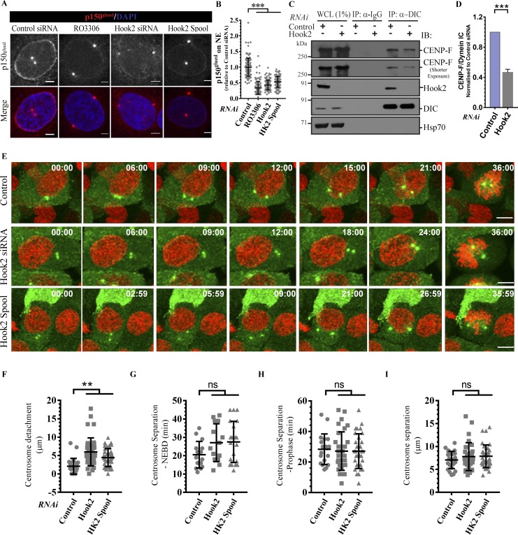
Figure 4.
Hook2 is required for p150glued localization to the NE during the late G2 phase by regulating CENP-F–dynein interaction. (A) Representative images showing loss of NE staining of p150glued upon Hook2 depletion. Nucleus was visualized by DAPI. Bars, 2 µm. (B) Quantification of the intensity of NE staining of dynactin in A (n = 3; 40 cells/experiment). (C) Lysates from HEK293T cells treated with indicated siRNA and synchronized to late G2 phase were incubated with IP with control IgG or anti-DIC antibody. The precipitates were IB with indicated antibodies. Hsp70 was used as a loading control. (D) Ratio of normalized band intensity (control siRNA) of IP CENP-F to DIC in C (n = 3). (E) Maximum intensity projections of z-stacks of live-cell time-lapse imaging of HeLa cells stably expressing EB1-GFP and H2B-mCherry and transfected with indicated siRNA. Cells were imaged every 3 m to monitor centrosome detachment and separation before mitotic entry. Bars, 10 µm. (F–I) Quantifications of centrosome detachment from the nucleus (F), the duration between the start of centrosome separation to NEBD (G) or prophase end (H), and the distance between centrosomes at prophase end (I) as measured from live-cell imaging experiments shown in E and analyzed from 30 cells. Data represent mean ± SD (ns, not significant; **, P < 0.01; ***, P < 0.001; Student’s t test).

