Figure 7.
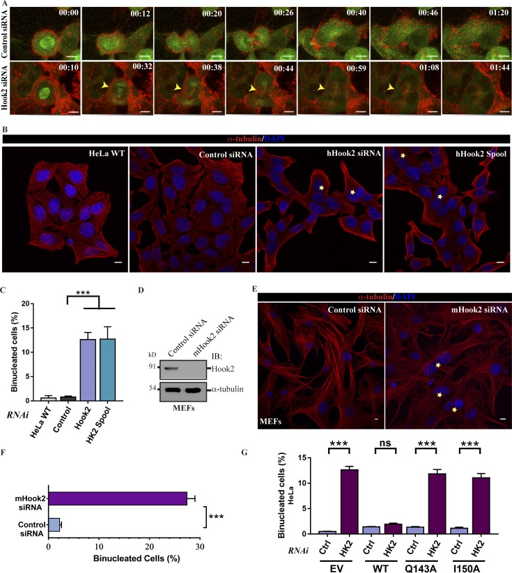
Hook2 depletion causes cytokinesis failure. (A) Maximum intensity projections of z-stacks of live-cell imaging of HeLa cells stably expressing GFP–α-tubulin and mCherry-UtrCH and treated with control or Hook2 siRNA. Z-stack time-lapse images were acquired every 3 m for a total duration of 3 h. Bars, 10 µm. (B) Representative images of HeLa cells treated with indicated siRNAs. The yellow asterisks in the images indicate binucleated cells in each case. Bars, 10 µm. (C) Quantification of binucleated HeLa cells as described in B (n = 5; 300 cells/experiment). (D) Western blot analysis confirming depletion of Hook2 in primary MEFs, and α-tubulin was used as a loading control. (E) Representative images of primary MEFs treated with control or mHook2 siRNA. The yellow asterisks in the images indicate binucleated cells in each case. Bars, 10 µm. (F) Quantification of binucleated primary MEFs as described in E (n = 5; 300 cells/experiment). (G) Quantification of the percentage of binucleated cells in siRNA-resistant Hook2 (WT/Q143A/I150A) transfected HeLa cells treated with control or Hook2 siRNA (n = 3; 200 cells/experiment). Data represent mean ± SD (ns, not significant; ***, P < 0.001; Student’s t test).

