Figure 1.
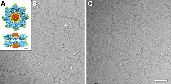
Cryo-electron micrographs of filamentous networks of RS1 molecules. (A) Architecture of the retinoschisin double rings, showing the N-terminal RS1 domains (red), the ring containing the disulfide bonds between subunits (yellow), the discoidin domain cores (blue), and the spikes (green; Tolun et al., 2016). Scale bar, 20 Å. (B and C) Two examples of micrographs of RS1 molecules forming branched networks of strands (arrows). The diamonds indicate four-way connections with a central RS1 molecule hub. The triangles indicate three-way connections. Scale bar, 500 Å.

