
Figure 2: Approximately one-third of all MCH neurons are CSF-projecting and these neurons are distinct from neuroendocrine neurons.
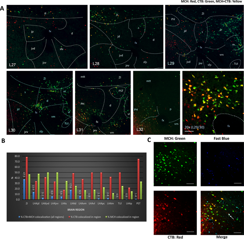
(A) Distribution of MCH immunolabeled and CTB retrogradely labeled neurons following lateral ventricular CTB injections; representative immunofluorescence for MCH (green), CTB (red), and colabeled cells (yellow). Numbers in the lower left in each image preceded by “L” indicate correspondence to closest atlas levels of the rat brain atlas of Swanson (Swanson, 2004). (B) Overall 32.7% of MCH-ir somata within the LHA and ZI were also CTB-ir, whereas 43.8 % of CTB-ir somata in the LHA and ZI were MCH-ir. Percentages of colabeling for the ZI and for specific LHA subregions are represented as follows: blue bars indicate the percentage of total (across all brain regions) colocalized MCH-ir + CTB-ir located in each region; red bars indicate the percentage CTB-ir neurons that are colocalized with MCH-ir in each region; green bars indicate the percentage MCH-ir neurons that are colocalized with CTB-ir in each region. (C) Representative images of immunofluorescence for MCH (green), CTB (red) and Fast Blue (FB, blue) in the lateral hypothalamic area. Intravenously injected FB retrogradely labelled neurons with access to the vasculature (putative neuroendocrine neurons). Overall 98.8% of MCH + CTB analyzed were not labeled with FB, with the arrow pointing to an extremely rare instance of a triple labeled cell (MCH + CTB + FB). Abbreviations: d = LHA dorsal region; fx = fornix; jd = LHA juxtadorsomedial region; jp = lateral hypothalamic area (LHA) juxtaparaventricular region; jvd = LHA juxtaventromedial region, dorsal zone; jvv = LHA juxtaventromedial region, ventral zone; m = LHA magnocellular nucleus; mtt = mammillothalamic tract; p = LHA posterior region; pc = LHA parvicellular region; PH = posterior hypothalamic nucleus; PST = preparasubthalamic nucleus; s = LHA suprafornical region; sfa = LHA subfornical region, anterior zone; sfp = LHA subfornical region, posterior zone; TUl = tuberal nucleus, lateral part; vl = LHA ventral region, lateral zone; vm = LHA medial zone, ventral region; ZI = zona incerta. Scale bar = 50 μm.