
Figure 6: MCH levels in the CSF are increased by DREADDs-mediated activation of MCH neurons and prior to nocturnal feeding.
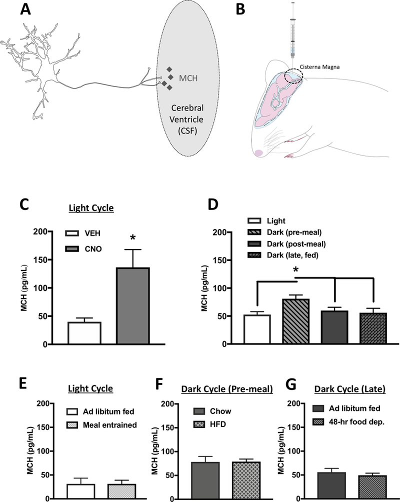
(A) A hypothetical model whereby MCH is transmitted into the CSF through axon terminals of ventricular-contacting terminals from MCH neurons. (B) Cartoon demonstrating the method of CSF extraction from the cisterna magna of an anesthetized rat. (C) MCH levels were elevated in CSF following MCH DREADDs activation (n=6,7). (D) Under physiological conditions, MCH levels in CSF are elevated during the early dark cycle prior to food consumption compared to during the light cycle and dark cycle postprandially (n=6–8). (E) There were no differences in CSF MCH levels prior to light cycle feeding in meal entrained animals compared to ad libitum fed controls (n=7/group). (F) Five days of exposure to a palatable high-fat diet had no effect on pre-prandial CSF MCH levels during the early dark cycle compared with chow-fed animals (n=6/group). (G) 48 hours of food deprivation had no effect on CSF MCH levels during the late dark cycle compared with ad libitum chow-fed controls. (n=5–6/group) (*P<.05). Data are means ± SEM.