Fig. 7.
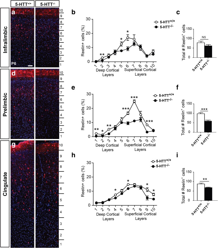
The number of reelin-positive cells is diminished in the absence of 5-HTT. Enlargements of cryosections of P6 5-HTT+/+ and 5-HTT−/− rat brains showing prefrontal swatches of the IL (a), PL (d), and the Cg (g) immunostained for reelin (red) and counterstained with DAPI (blue). Quantification of the percentage of reelin-positive neurons within the bins indicated in a, d, and g in the IL (b), PL (e), and Cg (h) of 5-HTT−/− compared to 5-HTT+/+ pups confirming the qualitative observations. Graphs in b, e, and h show average percentage of reelin-positive neurons normalized to total number of cells per bin ± SEM. One-way ANOVA, *p < 0.05, **p < 0.01, ***p < 0.001. Quantification of the total number of reelin-positive cells over the complete length of the prefrontal swatch in the IL (c), PL (f), and Cg (i) of 5-HTT−/− (black bar) compared to 5-HTT+/+ pups (white bar). Graphs in c, f, and i show average number ± SEM. One-way ANOVA, **p < 0.01, ***p < 0.001. Bar in a–g, 100 μm

