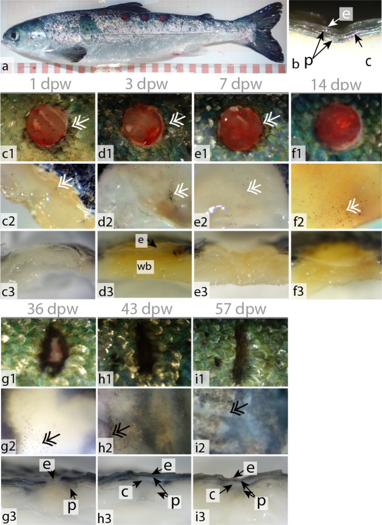
Figure 1.
Wound contraction and pigmentation, during the early (c–f) and late (g–i) healing phase. (a) Fish with three punch biopsy wounds. (b) Stereoscope picture of intact skin with the epidermal layer (e), pigment cells (p) and collagenous tissue (c). (c1–f1) Photographs of the wounds during the early healing phase. Pigmented bodies at the wound margins (double arrow). (c2–f2) Stereoscope pictures (40×) of the wound surface. (c3–f3) Stereoscope pictures (16×) of horizontally cut wounds showing the wound bed (wb). From 3 dpw and onward it is also possible to see the epidermal layer covering the entire wounded surface. (g1–i1) Photographs of the contracting wounds during the late healing phase. (g1–i1) Stereoscope pictures (40×) of the wound surface. (g3–i3) Stereoscope pictures (16×) of horizontally cut wounds showing the epidermal layer, pigment cells and collagenous tissue. Photographs N = 12, stereoscope pictures N = 6. Columns represent dpw and rows different orientation and magnification of the wound.

