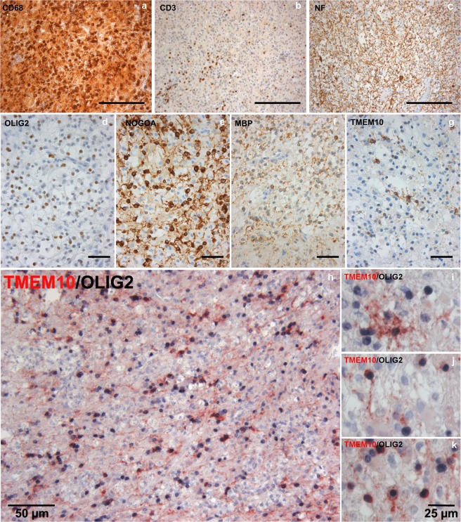
Figure 4.
TMEM10 is expressed by oligodendrocytes in remyelinating MS plaques –(a–f) Brain tissue samples containing an inflammatory demyelinating lesion consistent with MS were stained for CD68 and CD3 (markers of infiltrating immune cells), Neurofilament (axons), Olig2 (oligodendrocyte lineage cells), and TMEM10. NOGOA and MBP expression indicate ongoing remyelination in these plaques. (g) A subset of cells with oligodendroglial morphology expresses TMEM10. (h–k) Double staining for TMEM10 and Olig2 confirms that TMEM10 expressing cells are oligodendrocytes. Scale bars correspond to 200 µm in (a,b), 100 µm in (c), 50 µm in (d) through (h) and 25 µm.

