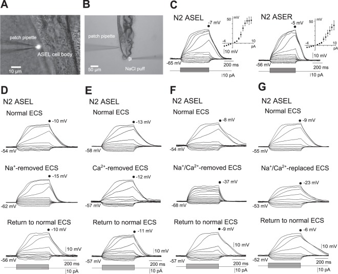
Figure 1.
Whole-cell, current-clamp recordings in vivo of ASE neurons in wild-type N2 animals. (A) Image of a patch-clamp recording. gcy-7p::GFP-transfected N2 specifically producing GFP in ASEL was glued to a cover glass, and was transferred to a recording chamber. Under a microscope, a small piece of cuticle and body wall was dissected to exteriorize ASEL cell bodies. (B) Image of an animal under stimulation with NaCl-containing buffer. Animals were treated with the same procedures as in (A), and their nose tips were stimulated with NaCl solutions released from a puffing capillary. (C) Membrane voltage changes in response to current steps and averaged I-V curves in wild-type ASEL (left, n = 22) and ASER (right, n = 7). Error bars, s.e.m. (D–G) Membrane voltage changes in response to current steps in wild-type ASEL. Current-clamp recordings of an animal were carried out three times consecutively, first and third in normal ECS (top and bottom), and the second in ECS, from which Na+ was removed (Na+-removed ECS) (D), Ca2+ of which was removed (Ca2+-removed ECS) (E), Na+ and Ca2+ of which were removed (Na+/Ca2+-removed ECS. In this experiment, current was injected until the membrane potential exceeded −40 mV) (F), or Na+ and Ca2+ of which were replaced with NMDG+ and Mg2+ (Na+/Ca2+-replaced ECS) (G). These are representatives of at least three independent recordings.

