Figure 3.
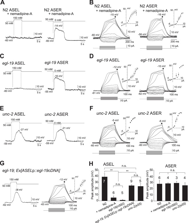
EGL-19 plays an essential role in active propagation of electrical signals in ASEL. (A,B) Membrane voltage traces of wild-type ASEL and ASER in the presence of nemadipine-A, 1.0 μM, upon stimulation with 150 mM NaCl and NaCl-free buffer, respectively, to the nose tips of animals (A), or upon current steps with mean I-V curves (ASEL: n = 6, ASER: n = 4) (B). (C,D) Membrane voltage traces of ASEL and ASER in egl-19 mutants upon stimulation with 150 mM NaCl and NaCl-free buffer, respectively (C), or upon current injection with mean I-V curves (ASEL: n = 5, ASER: n = 3) (D). (E,F) Membrane voltage traces of ASEL and ASER in unc-2 mutants upon stimulation with 150 mM NaCl and NaCl-free buffer, respectively (E), or upon current injection with mean I-V curves (ASEL: n = 5, ASER: n = 4) (F). (G) Membrane voltage traces of ASEL in egl-19 transgenic animals specifically producing wild-type EGL-19 in ASEL upon stimulation with 150 mM NaCl buffer (left), or upon current injection with mean I-V curves (right, n = 9). (H) Mean amplitudes of membrane voltage peaks of the traces shown above in (A,C,E,G). Numbers above bars indicate numbers of animals measured. N2 data are derived from Fig. 2D. **p < 0.01, n.s., not significant by one-way ANOVA with Dunn’s test. Error bars, s.e.m.

