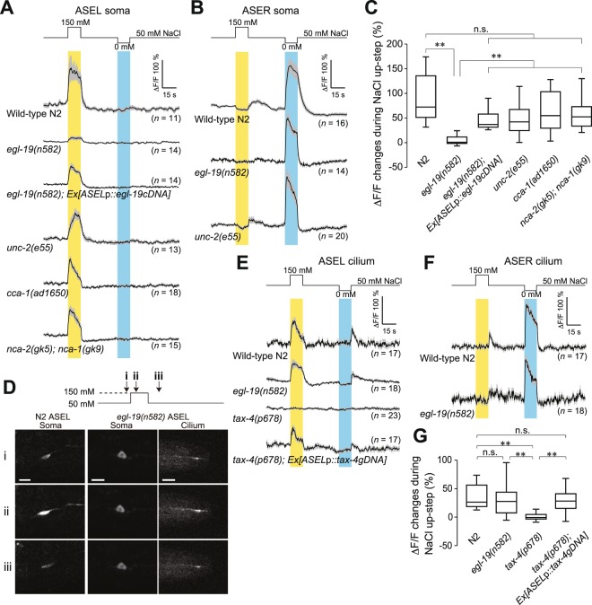
Figure 4.
Ca2+ transients in cell body and sensory cilium of ASE neurons of animals immobilized in microfluidic chambers. (A) Ca2+ transients in ASEL cell bodies of wild type, egl-19, ASEL-specifically rescued egl-19, unc-2, cca-1, and a double mutant, nca-2; nca-1, in response to NaCl concentration changes for 15 s, which are shown on top. Grey bands, s.e.m. (B) Ca2+ transients in ASER cell bodies of wild-type, egl-19, and unc-2 animals in response to NaCl concentration changes for 15 s. (C) Mean ΔF/F changes in ASEL cell bodies measured in (A) during 15-s stimulation with 150 mM NaCl buffers. Horizontal lines in boxes indicate 25th, 50th, and 75th percentiles, and whiskers represent 5th and 95th percentiles. (D) Upon stimulation of ASEL cilia with 150 mM NaCl buffer for 15 s, Ca2+ transients of ASEL cilia of egl-19(n582) were monitored 3 s before the stimulation (i), 5 s after the NaCl up-step (ii), and 8 s after cessation of the stimulation (iii). Note that Ca2+ influx into the cilia, but not to the cell body, of egl-19 ASEL was detected. Ca2+ transients in the ASEL cell body of wild-type N2 are also shown as references at the left. (E) Ca2+ transients in ASEL cilia of wild type, egl-19, tax-4, and tax-4 rescued by ASEL-specific expression of tax-4 genomic DNA, in response to NaCl concentration changes for 15 s as shown on top. (F) Ca2+ transients in ASER cilia of wild-type and egl-19 animals in response to NaCl concentration changes for 15 s. (G) Mean ΔF/F changes in ASEL cilium in (E) during 15-s stimulation with 150 mM NaCl buffers. Horizontal lines in boxes indicate 25th, 50th, and 75th percentiles, and whiskers represent 5th and 95th percentiles. **p < 0.01, n.s., not significant by Steel-Dwass test.

