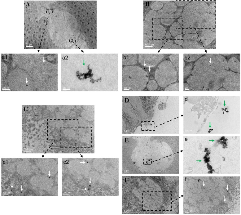
Fig. 4.
Distribution of triangular silver nanoplates in intestinal GCs of mice with CBD ligation. The CBD ligation mice group was treated with triangular silver nanoplates injected via tail vein 7 days after ligation. Intestinal GCs of different intestinal tissues. A Duodenum, triangular silver nanoplates were shown in an aggregation mode (green arrow), while some triangular sliver nanoplates were in a dispersion mode (white arrows). B Jejunum, triangular silver nanoplates located at the intestinal GC (white arrows). C Ileum and some triangular silver nanoplates were excreted out, while some were still inside. D Colon, some triangular silver nanoplates were secreted out and into the gut. E, F Rectum, some triangular silver nanoplates were ready to excrete out (dispersion mode, white arrows), while others were still inside (aggregation mode, white arrow)

