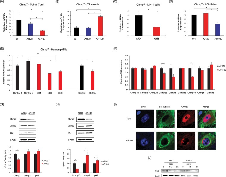
Figure 2.
Chmp7 is differentially regulated in in presymptomatic SBMA mice and motor neuron precursor cells derived from SBMA patients. (A) Chmp7 was downregulated in AR100 mice spinal cord compared to control WT and AR20 presymptomatic male mice at 3 months of age. (B) Chmp7 expression was upregulated in tibialis anterior (TA) muscle of presymptomatic AR100. (C) Chmp7 was also downregulated in the SBMA AR65Q neuronal stable cell line model and (D) in laser captured (LCM) ventral spinal cord motor neurons from presymptomatic AR100 mice. (E) Chmp2b was downregulated in AR100 mice, while Chmp4c was increased. qPCR data are displayed as mean ± SEM and are representative of at least three independent experiments. Statistical analysis was performed using a two sample t-test or one-way ANOVA followed by the Student–Newman–Keuls and Tukey’s Honestly Significantly Different post hoc tests (n ≥ 3, *P < 0.05). (E) CHMP7 was downregulated in SBMA patient-derived motor neuron precursor cells (pMN). (*P < 0.05, **P < 0.01, two sample t-test or ANOVA with Tukey’s Honestly Significantly Different post hoc test). (G) The protein levels of Chmp7, Lamp2 and p62 were analysed by Western blot of spinal cord of AR100 and control AR20 male mice. Chmp7 was decreased in presymptomatic (3 month) AR100 mice. (H) Chmp7 was further reduced in symptomatic (12 month) AR100 mice, while Lamp2 levels were increased. Densitometric analysis of bands was performed using values normalized to actin. Data are displayed as mean ± SEM and are representative of three independent experiments. Statistical analysis was performed using a two sample t-test (n ≥ 3, *P < 0.05). AU = arbitrary units. (I) CHMP7 staining in motor neurons of 3 month old presymptomatic WT and AR100 mice. β-III tubulin was used to stain motor neurons. Scale bars represent 20 μm. (J) TrkB degradation was performed using primary motor neuron cultures starved for 1 h in serum-free medium without BDNF. Cells were incubated with BDNF and cycloheximide (CHX). In AR100 cultures there was a delay in the degradation of TrkB compared to WT.

