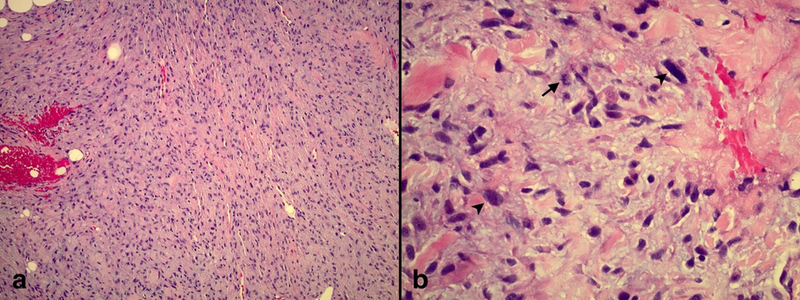
Figure 3.
Punch biopsy of scalp lesion. a) Spindled cells arranged in sheets and fascicles within a myxoid stroma with focal invasion into the dermal fat (H&E, 10X magnification). b) Tumor cells have pale eosinophilic cytoplasm and wavy, tapered nuclei with nuclear atypia (arrowhead) and mitoses (arrow) (H&E, 40X magnification).

