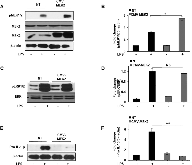
FIGURE 6. MEK2 overexpression decreases IL-1β production with no significant effect on ERK activation.

RAW264.7 cells were transfected with a Mek2-Myc construct (CMV-MEK2) for 24h or cultured without plasmid (NT). MEK2 overexpressed or NT cells were treated with LPS (100 ng/mL) for 30 minutes or 3h. (A) Whole cell lysates were subjected to SDS-PAGE followed by Western blot analysis using antibodies against MEK2 and phospho-specific antibody against MEK1/2. Equal loading was determined using antibody against β-actin. (B) Densitometric values expressed as fold change of the ratio pMEK1/2/β-actin. (C) Western blot analysis was performed using antibodies against phospho-specific ERK1/2 and total ERK. (D) Densitometric values expressed as fold change of the ratio pERK1/2/total ERK. (E) Western blot analysis was performed using antibody against pro IL-1β, equal loading was determined using antibody against β-actin. (F) Densitometric values expressed as fold increase of the ratio pro IL-1β/β-actin. Data presented for all experiments are representative of at least 3 independent experiments. Using ANOVA Mann-Whitney U test for all results, a p value <0.05 was considered significant and error bars indicate SEM.
