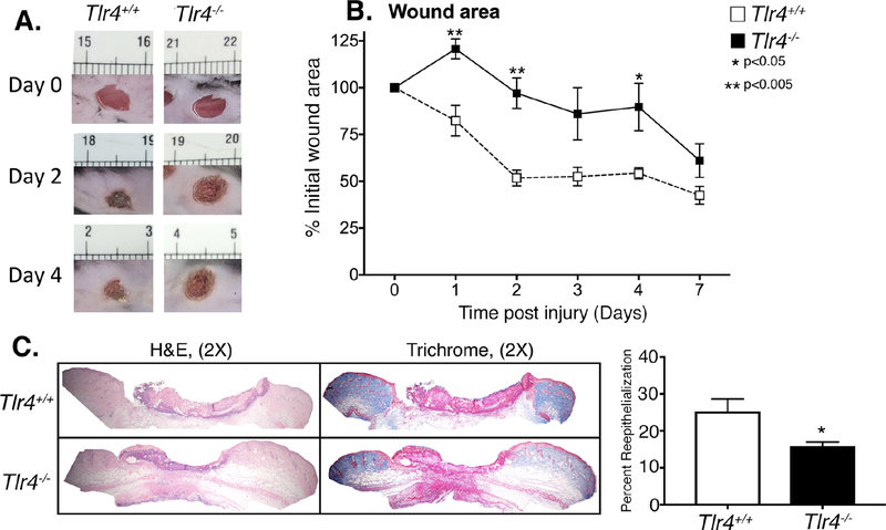
Figure 2. TLR4 deficient mice exhibit delayed wound healing and decreased reepithelization.
A: Wounds were created in Tlr4−/− and control mice. Representative photographs of the wounds of Tlr4−/− mice and controls on days 0 and 4 post injury are shown. B: The change in wound area was recorded daily by blinded observer and analyzed with ImageJ software (n = 5). C: Wounds were harvested on day 3, paraffin embedded and sectioned. 5 μM sections were stained with hematoxylin and eosin and with Masson’s Trichrome stain. Percent re-epithelialization was calculated by measuring distance traveled by epithelial tongues on both sides of wound divided by total distance for full re-epithelialization. Representative images are shown in 2X magnification (*P < 0.05; n=5; repeated 1X). Data are presented as the mean±SEM. Data are representative of 2–3 independent experiments. Data were first analyzed for normal distribution and if data passed normality test, 2-tailed Student t test was used.

