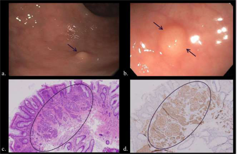
Figure 14.

Two patients undergoing colonoscopy for history of colon polyps (a, b) and iron deficiency anemia (c. d), respectively. a, b) Small sub-mucosal sessile polypoid lesions (arrows) were removed with excisional biopsy. c, d). Hematoxylin and eosin (c) microscopy demonstrates cellular clusters of neuroendocrine cells in the submucosa and lamina propria (circled) that stain for synaptophysin (d).
