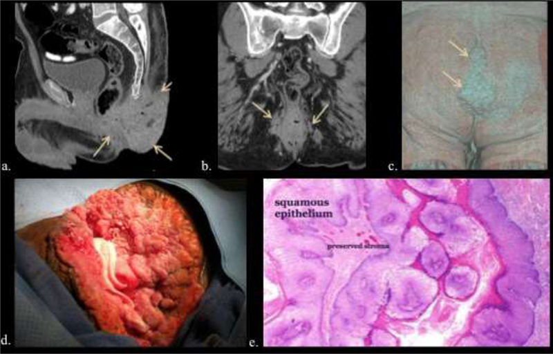
Figure 16.

49-year-old male with 14-year history of increasing anal warts. a, b, c) Multi- planar CT (a, b) and volume rendered image (c) depict extensive lobulated anal and perianal disease extending from the gluteal cleft (arrows). d, e). Intraoperative image of giant condylomata (d) and low power microscopy of a typical non-invasive wart (e).
