Figure 2.

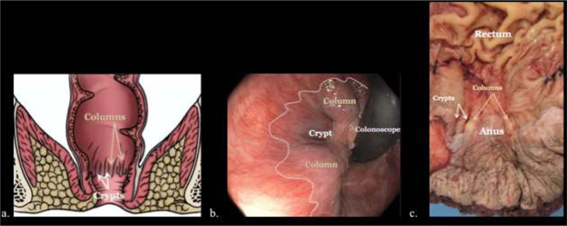
Illustrative diagram (a), endoscopic image (b), and gross pathologic specimen (c) demonstrating the columns of Morgagni (gold arrows, a, c), anal crypts (white arrows, a, c) between the columns and dentate line (dashed line, b).

Illustrative diagram (a), endoscopic image (b), and gross pathologic specimen (c) demonstrating the columns of Morgagni (gold arrows, a, c), anal crypts (white arrows, a, c) between the columns and dentate line (dashed line, b).