Fig. 3.
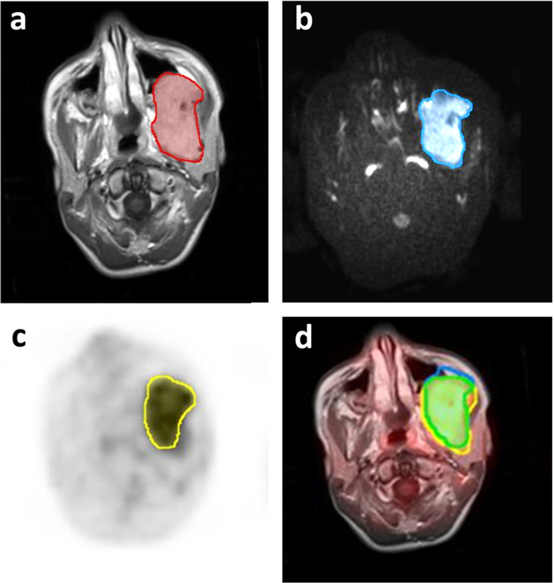
[18F]FDG PET and MR scans of a 6-year-old girl with left nasopharyngeal embryonal rhabdomyosarcoma before start of therapy. a Manually drawn region of interest (ROI) outlines the whole tumor mass on contrast-enhanced T1 weighted image. b ROI outlines tumor mass with restricted diffusion. c ROI outlines tumor mass with increased [18F]FDG metabolic activity on PET. d Eighty-eight percent of the tumor tissue showed an overlap of [18F]FDG metabolic activity and restricted diffusion. The patient demonstrated progressive disease at 2.4 years after therapy.

