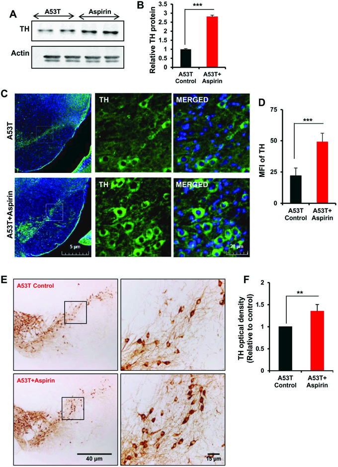
Figure 4. Oral administration of aspirin increases the level of TH in vivo in the nigra of A53T-Tg mice.

Male A53T-Tg mice (n=5 per group) were treated with aspirin (2 mg/kg body wt/d) mixed in 0.5% methylcellulose orally via gavage. Control mice received 0.5% methylcellulose as vehicle. After 30 d of treatment, the level of TH was monitored in the SNpc by Western blot (A). Actin was run as loading control. Bands were scanned and values (TH/actin) presented as relative to control (B). Results are mean + SEM of five mice per group. ***p<0.001 (= 1.865 × 10⁻⁴) vs control by two-tailed paired t-tests. The level of TH was monitored in ventral midbrain sections by immunofluorescence (C). MFI of TH (D) was calculated in two nigral sections of each of five mice per group. ***p<0.001 (= 4.69 × 10⁻⁹) vs control by two-tailed paired t-tests. The level of TH was monitored in ventral midbrain sections by DAB immunostaining (E). Optical density of TH (F) was calculated in two nigral sections of each of five A53T-Tg mice per group. Results are mean ± SEM of five mice per group. **p<0.01 (= 0.00245) vs control by two-tailed paired t-tests.
