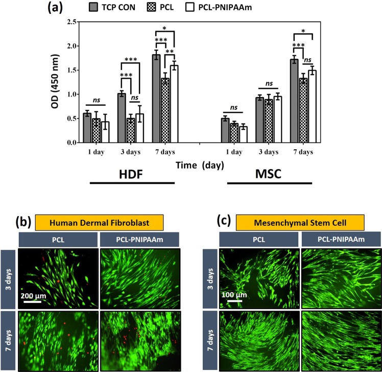
Figure 3.
Cell proliferation on PCL and PCL-PNIPAAm surfaces. CCK-8 studies showed that both HDF cells and MSCs survived and proliferated on both PCL and PCL-P (PCL-PNIPAAm) using tissue culture plates as control (TCP CON) surfaces for 1, 3 and 7 days (a). At day 7, a significant increase in OD was observed in both cell types compared to day 1 and 3 and both types proliferated more on the surfaces of PCL-P than the surfaces of PCL (*p < 0.05; **p < 0.01; ***p < 0.001; ns: no significant difference). This trend of cell viability and proliferation was also observed in fluorescent microscopy for both HDF cells (b) and MSCs (c). HDF cells were stained with Live-dead staining (green: membrane of living cells, red: nuclei of dead cells) whereas GFP was cloned into MSCs. Emission was observed with fluorescent microscopy at days 3 and 7. Very dense and clustered cells with higher viability were observed at day 7 and on modified surfaces (PCL-P) than at day 3 and on non- modified PCL surfaces.

