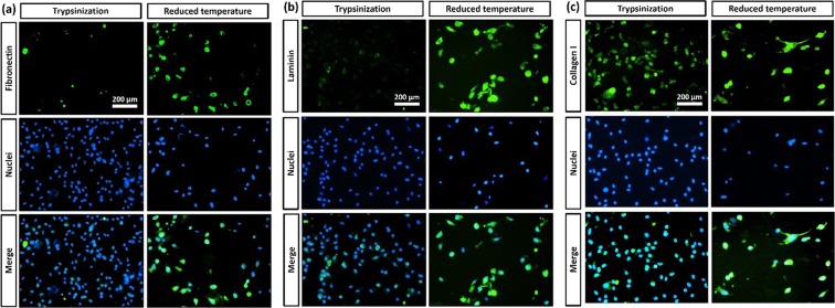
Figure 6.
Immunofluorescent analysis. Representative Immunostaining results for ECM proteins of HDF cells. (a) Fibronectin, (b) Laminin and (c) Collagen I on cells detached from PCL-PNIPAAm macrocarriers by trypsin treatment and reduced temperature. Nuclei stained blue with Hoechst 33342 dye and proteins stained green with FITC-labelled secondary antibody. Immunofluorescent images showed that cells detached by reduced temperature contained fibronectin whereas no fibronectin was found in cells detached by trypsin treatment.

