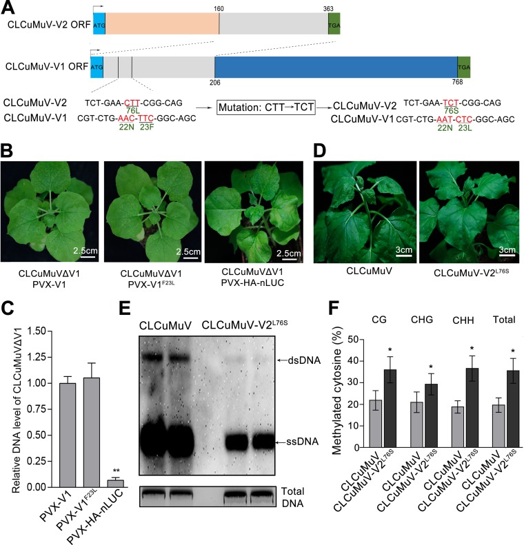
FIG 7.
Replacement of V2 with V2L76S attenuates CLCuMuV infection. (A) The V2 open reading frame (ORF) (nt 160 to 365) overlaps with the V1 ORF (nt 1 to 206) in the CLCuMuV genome. Thus, an L76S mutation in V2 (CTT-TCT) caused the F23L mutation in V1. (B) N. benthamiana was coinfected with CLCuMuVΔV1 and PVX-V1, PVX-V1F23L, or PVX-HA-nLUC. Photographs were taken under white light at 14 dpi. (C) qPCR showing the CLCuMuV DNA levels in the systemic leaves of plants coinfected with CLCuMuVΔV1 and PVX-V1, PVX-V1F23L, or PVX-HA-nLUC. Data were obtained from three independent experiments. The asterisks indicate significant differences (Student’s t test, P < 0.01). The bars represent mean ± SD. (D) Plants infected with CLCuMuV-V2L76S, which contains an L76S mutation in V2, showed weaker viral symptoms than those infected with CLCuMuV. Photographs were taken at 16 dpi. (E) Southern blot assay showing DNA accumulation for CLCuMuV and CLCuMuV-V2L76S. A partial CLCuMuV V1 DNA was used as a template to make a 32P-labeled probe. Single-stranded viral DNA (ssDNA) and double-stranded viral DNA (dsDNA) DNA are indicated by arrows. Total genomic DNA was stained with ethidium bromide as a loading control. (F) Detection of the cytosine methylation status of the viral 5′ intergenic region (IR) in CLCuMuV- or CLCuMuV-V2L76S-infected plants. The histogram shows the proportions of methylated cytosine residues in different sequence groups. The asterisk indicates significant differences (*, P < 0.05, Student’s t test). The bars represent means ± SD. Data were obtained from three independent experiments.

