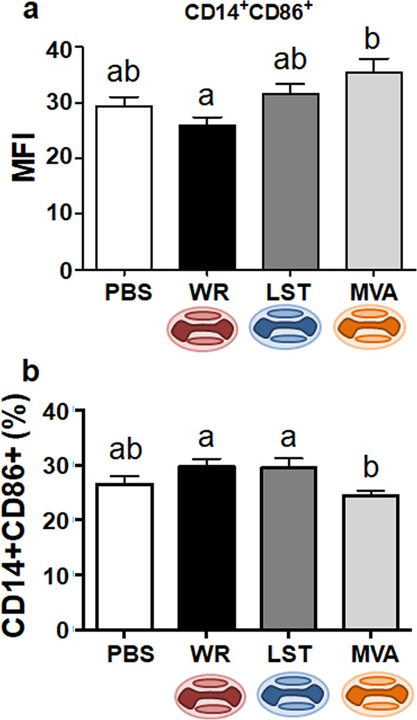
FIG 3.

Expression of the CD86 activation marker in monocytes/macrophages from VACV-infected or uninfected mice. Splenocytes from mice euthanized at 14 days p.i. were incubated with fluorochrome-labeled antibodies to detect the expression of CD14 and CD86 molecules on cell surfaces by flow cytometry. Splenocytes were from intranasally (a) or intraperitoneally (b) infected mice. Bars represent the means ± standard errors of the mean fluorescence intensity (MFI) values of costimulatory molecules on macrophages (a) or the means ± standard errors of each cell phenotype frequency within the gated population (b). Different letters indicate statistical differences (P < 0.05) between groups (n = 7). For better visualization and orientation, colorful virus cartoons represent virus-infected groups in all figures (red, VACV-WR; blue, VACV-LST; orange, MVA).
