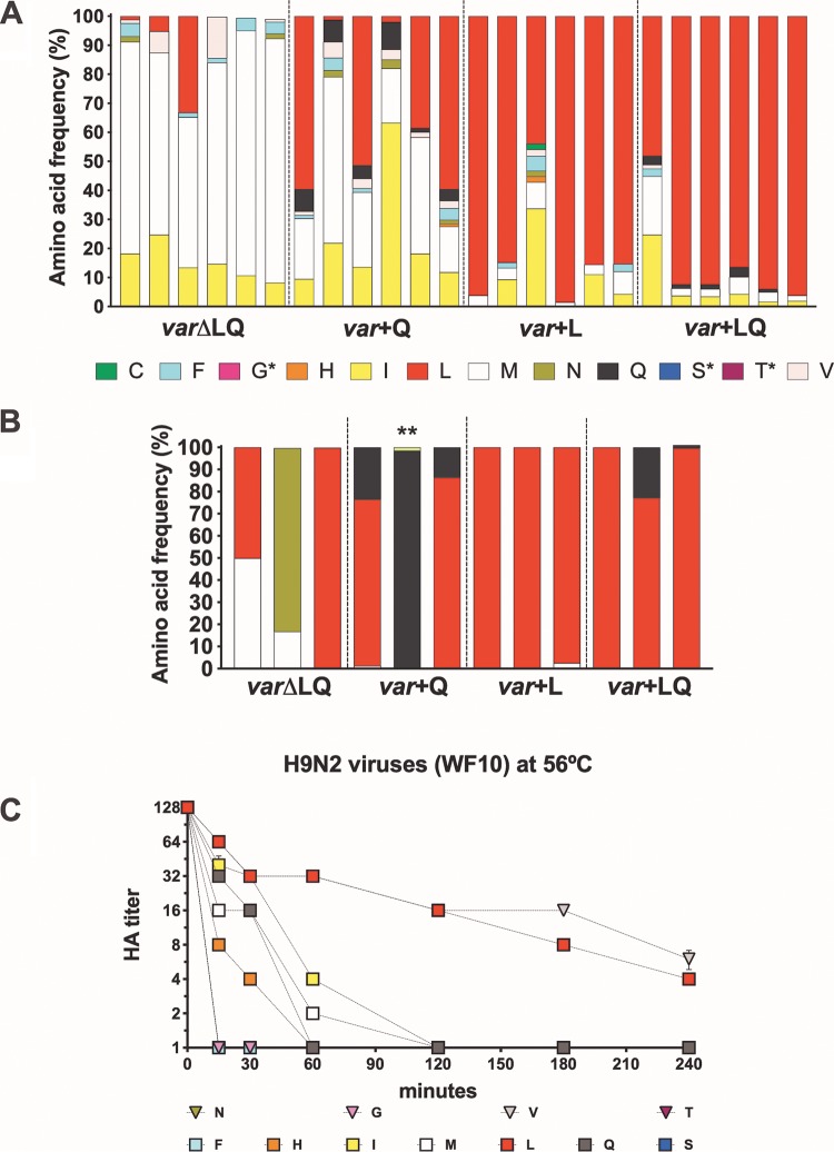
FIG 8.
Frequency of amino acids present in tracheal swab samples of inoculated and contact quails and temperature stability of H9N2 variant viruses. The viruses in tracheal swab specimens collected at 3 dpi from inoculated quails (A) and 8 dpc from contact quails (B) were sequenced by NGS, and the amino acid frequency was analyzed using Geneious (version 10.2.3) software. Each bar represents a quail in the respective group, and each color represents a variant virus. Single asterisks refer to variant viruses included in the virus mix and not identified in tracheal swabs, while the double asterisk shows the K226 variant virus not included in the virus mix yet identified in contact quail. (C) Temperature stability of H9N2 variant viruses at 56°C. Variant viruses diluted to 128 HAU/50 μl were incubated at 56°C. Samples collected at 0, 15, 30, 60, 120, 180, and 240 min postincubation were used for hemagglutination assays. Treatment was carried out in quadruplicate. L, Q, M, F, I, S, H, N, V, C, T, and G correspond to the amino acid mutations at position 226 in the HA of H9N2 viruses. Naturally occurring amino acids previously found in H9 HA are shown as squares; others are shown as triangles. Differences were observed depending on which amino acid was present at position 226. By 1 h at 56°C, all H9N2 viruses had hemagglutination titers (HA titers) of less than 2 HAU, except viruses with aliphatic amino acids, V226, L226, and I226 viruses, as well as the C226 and Q226 viruses. After 4 h, only the viruses with V226 and L226 showed any hemagglutination titer and were the most stable viruses.

